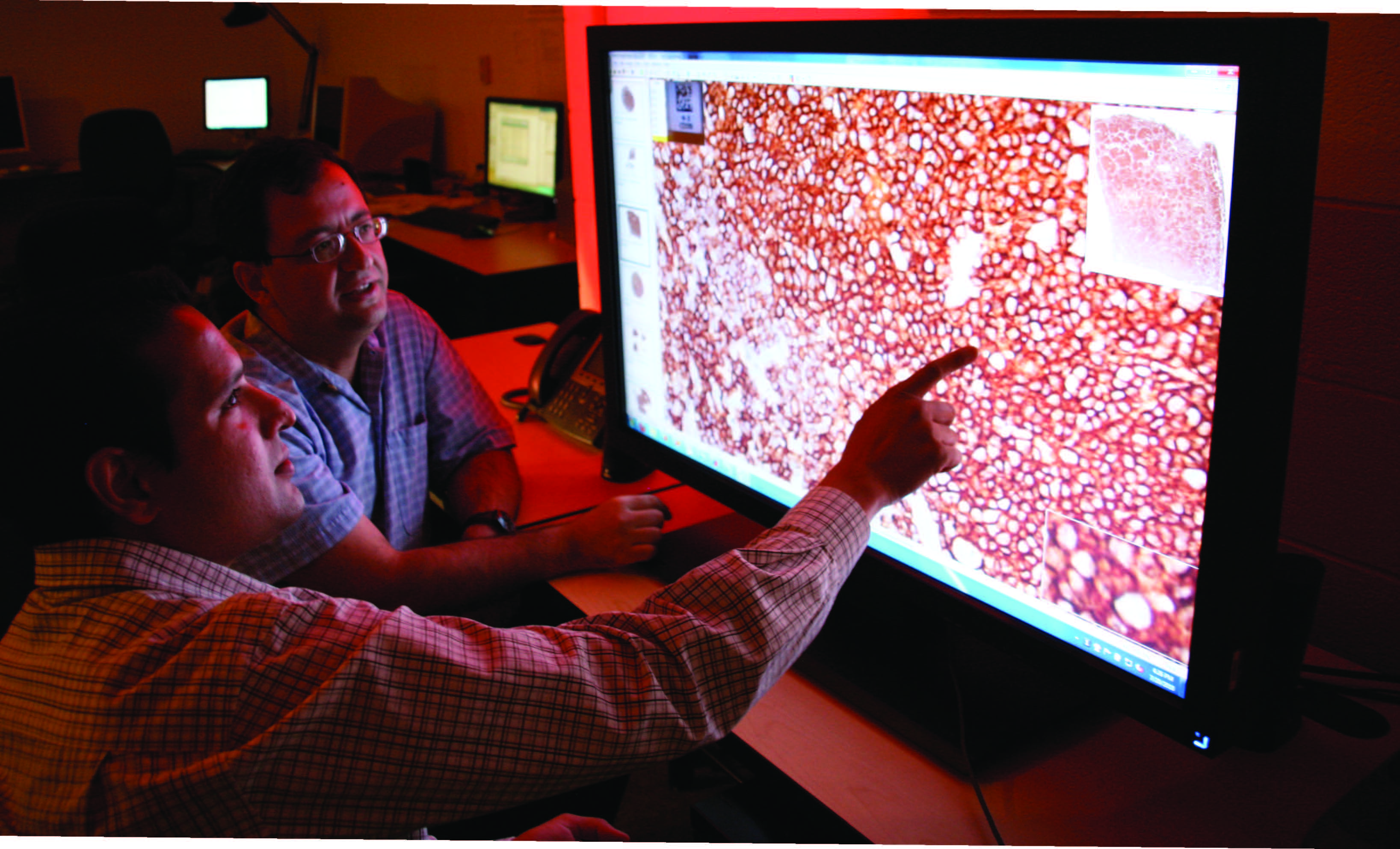
above: Metin Gurcan discusses a feature Siddharth Samsi has found on a digitized slide specimen being examined for indications of Follicular Lymphoma. Their Ohio State University Medical Center research group has been leveraging Ohio Supercomputer Center resources to develop a computer-assisted diagnosis tool to improve grading of the common, slow-growing cancer.
Follicular Lymphoma (FL) is one of the most common forms of non-Hodgkin Lymphoma occurring in the United States. FL is a slow-growing cancer of the human lymph system that usually spreads into the blood, bone marrow and, eventually, internal organs. A World Health Organization pathological grading system is applied to a biopsy sample; doctors usually avoid prescribing severe therapies for lower grades, while they usually recommend radiation and chemotherapy regimens for more aggressive grades.
Accurate grading of the pathological samples generally leads to a promising prognosis, but diagnosis depends solely upon a labor-intensive process that can be affected by human factors such as fatigue, reader variation and bias. Pathologists must visually examine and grade the specimens through high-powered microscopes. Now, computer-assisted diagnosis tools are being developed to provide pathologists with higher throughput of specimen analyses and, ultimately, quicker, accurate diagnoses.
“The advent of digital whole-slide scanners in recent years has spurred a revolution in imaging technology for histopathology,” according to Metin N. Gurcan, Ph.D., an assistant professor of biomedical informatics at The Ohio State University Medical Center. “The large multi-gigapixel images produced by these scanners contain a wealth of information potentially useful for computer-assisted disease diagnosis, grading and prognosis.”
Processing and analysis of such high-resolution images, Gurcan points out, remain non-trivial tasks, not just because of the sheer size of the images but also due to complexities of underlying factors involving differences in staining, illumination, instrumentation and goals. To overcome many of these obstacles to automation, Gurcan and medical center colleagues, Dr. Gerard Lozanski and Dr. Arwa Shana’ah, turned to the Ohio Supercomputer Center. Ashok Krishnamurthy, Ph.D., interim co-executive director of the center, and Siddharth Samsi, a computational science researcher there and an OSU graduate student in Electrical and Computer Engineering, put the power of a supercomputer behind the process.
“Our group has been developing tools for grading of follicular lymphoma with promising results,” said Samsi. “We developed a new automated method for detecting lymph follicles using stained tissue by analyzing the morphological and textural features of the images, mimicking the process that a human expert might use to identify follicle regions. Using these results, we developed models to describe tissue histology for classification of FL grades.”
Histological grading of FL is based on the number of large malignant cells counted in within tissue samples measuring just 0.159 square millimeters and taken from ten different locations. Based on these findings, FL is assigned to one of three increasing grades of malignancy: Grade I (0-5 cells), Grade II (6-15 cells) and Grade III (more than 15 cells).
“The first step involves identifying potentially malignant regions by combining color and texture features,” Samsi explained. “The second step applies an iterative watershed algorithm to separate merged regions and the final step involves eliminating false positives.”
The large data sizes and complexity of the algorithms led Gurcan and Samsi to leverage the parallel computing resources of OSC’s Glenn Cluster in order to reduce the time required to process the images. They used MATLAB. and the Parallel Computing Toolbox™ to achieve significant speed-ups.
Speed is the goal of the research project, but accuracy is essential. Gurcan and Samsi compared their computer segmentation results with manual segmentation and found an average similarity score of 87.11 percent.
“This algorithm is the first crucial step in a computer-aided grading system for Follicular Lymphoma,” Gurcan said. “By identifying all the follicles in a digitized image, we can use the entire tissue section for grading of the disease, thus providing experts with another tool that can help improve the accuracy and speed of the diagnosis.”
--
Project lead: Metin N. Gurcan, The Ohio State University
Research title: Computer-aided analysis of immunohistochemical slides of follicular lymphoma
Funding source: National Cancer Institute