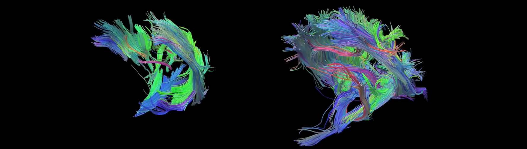
Streamline tractography visualizations for a study investigating the use of a form of vitamin E to reduce tissue damage inflicted by stroke.
Researchers at The Ohio State University’s Center for Clinical and Translational Science (CCTS) are using sophisticated scanners and powerful supercomputers to study how vitamin E can be used to reduce the extent of brain injury suffered by stroke patients.
Cameron Rink, Ph.D., assistant professor of surgery in the College of Medicine at Ohio State’s Wexner Medical Center, is part of a research team that has been investigating stroke treatments for more than a decade. He teamed up with a pair of computer scientists who leveraged Ohio Supercomputer Center resources to create detailed visualizations of brain activity during a stroke, providing researchers with an extraordinary window into treatments for ischemia, the loss of blood flow and resulting tissue damage.
Supported by CCTS funding, Rink discovered that the preventive use of a form of vitamin E called tocotrienol reduces stroke-induced brain damage in small animals in multiple ways. Tocotrienol appears to enhance the body’s natural ability to use other blood vessels in the brainto bypass a clot and may reduce the chances of a repeat stroke.
“The study involved combining MR diffusion tensor imaging with streamline tractography techniques,” explained Raghu Machiraju, Ph.D., an associate professor in both computer science and engineering and biomedical informatics at Ohio State University. “Originally, Dr. Rink was doing this himself on a laptop. We were able to apply some sophisticated preprocessing tools to the data and access OSC’s ‘big iron’ to do the analysis. It’s computationally very expensive.”
In neuroscience, streamline tractography is a procedure that illustrates an implied flow along neural pathways, or ‘tracts,’ using special magnetic resonance imaging (MRI) techniques and computer-based image analysis to produce two- and three-dimensional visualizations. This study used diffusion tensor MRI (DTI) imaging of an animal’s brain both during and after a stroke. This allowed researchers to glean tracts of white matter to determine the damage to the surrounding tissue.
Machiraju and Okan Irfanoglu, Ph.D., then an OSU research assistant, ran more than 60,000jobs to apply a complex set of algorithms to large data files from seven MRI scans to create the visualizations. Irfanoglu now works at the National Institute of Child Health and Human Development of the National Institutes of Health in the lab of Peter Basser, Ph.D., and Carlo Pierpaol, M.D., Ph.D., the inventors of DTI.
---
Project lead: Raghu Machiraju, The Ohio State University
Research title: Tocotrienol vitamin E protects against preclinical canine ischemic stroke by inducing arteriogenesis
Funding source: The Ohio State University Center for Clinical and Translational Science
Website: http://bit.ly/OSC-RR-Machiraju